
|
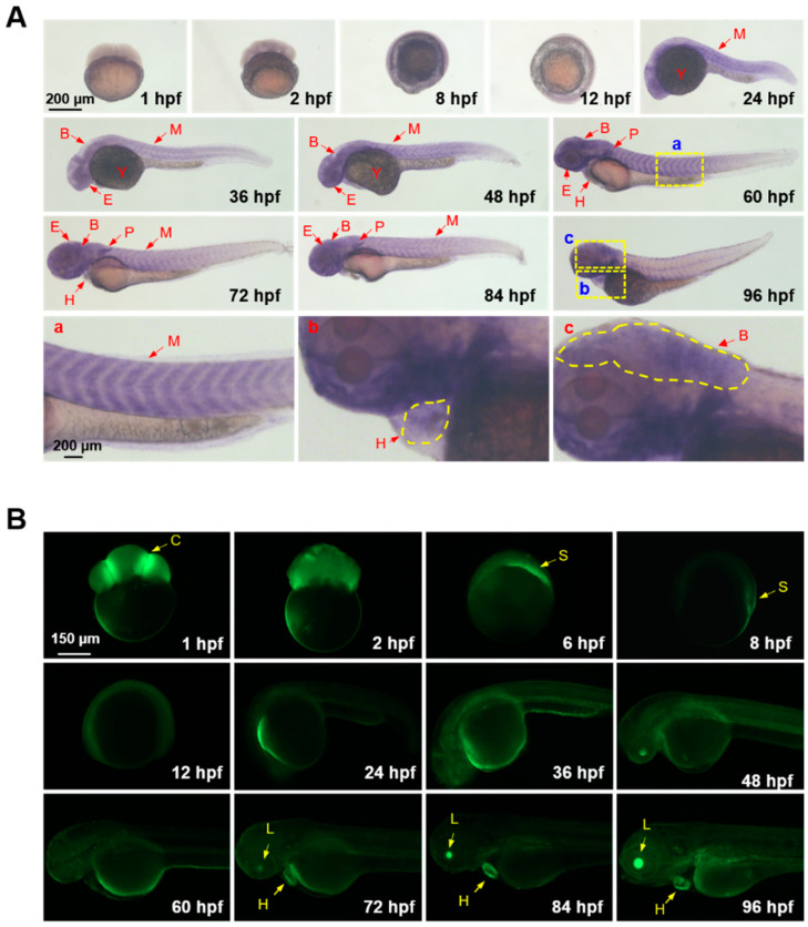
Figure 2 GCaMP6s expression and Ca2+ signaling activity in embryos and larvae of the transgenic zebrafish. (A) Spatiotemporal expression patterns of GCaMP6s analyzed by whole-mount in situ hybridization. The magnified images indicate the expression of GCaMP6s in the myotome (a), heart (b) and brain (c). (B) Fluorescent microscope images indicating Ca2+ signaling activities in the anatomical structures of zebrafish embryos and larvae. Lateral view of the early embryos (before 12 hpf) are shown. The embryos and larvae after 24 hpf were oriented as head to the left and dorsal to the top. B, brain; C, cleavage furrow; E, eyes; H, heart; L, lens; M, myotome; P, pectoral fin; S, embryonic shield; Y, yolk sac.