
IMAGE
Figure 7
- ID
- ZDB-IMAGE-210611-64
- Antibodies
- Publication
- Crouzier et al., 2021 - Loss of Pde6a Induces Rod Outer Segment Shrinkage and Visual Alterations in pde6aQ70X Mutant Zebrafish, a Relevant Model of Retinal Dystrophy
- All Figures
- Figures for Crouzier et al., 2021
Image
|
Figure Caption
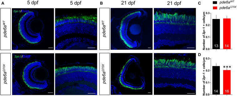
Figure 7
Analysis of cones in
Figure Data
Acknowledgments
This image is the copyrighted work of the attributed author or publisher, and
ZFIN has permission only to display this image to its users.
Additional permissions should be obtained from the applicable author or publisher of the image.
Full text @ Front Cell Dev Biol