
FIGURE 7
- ID
- ZDB-IMAGE-210611-57
- Genes
- Publication
- Morris et al., 2021 - A Novel Lysolecithin Model for Visualizing Damage in vivo in the Larval Zebrafish Spinal Cord
- All Figures
- Figures for Morris et al., 2021
|
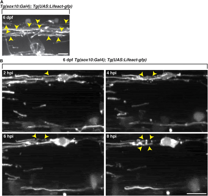
FIGURE 7
Oligodendrocyte cytoskeletal components dynamically change following exposure to lysolecithin. All images are lateral views of the spinal cord with anterior to the left and dorsal to the top.