IMAGE

Figure 5.3

ID
ZDB-IMAGE-210607-27
Source
Figures for Okuda et al., 2021
Image
Figure Caption

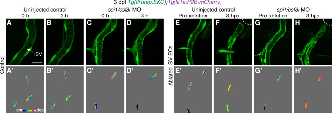
Figure 5.3 Macrophages are not required to sustain Erk activity in ablated ISV ECs following vessel wounding.

(A–H’) Lateral confocal images of ISV endothelial cells (ECs) in 3 days post-fertilisation (dpf) EC-EKC uninjected control (A–B’, E–F’) and spi1/csf3r morphants (C–D’, G–H’). Images (A-D’) show non-ablated control ISV ECs. Images (E) and (G) were taken pre-ablation and images (F) and (H) were taken 3 hours post-ablation (hpa). Images (A-H) show fli1aep:EKC expression, and images (A’-H’) show the nuclear fli1aep:EKC intensity. White dotted lines show the wounded site of each embryo. ISV: intersegmental vessel. Scale bar: 20 μm.

Acknowledgments
This image is the copyrighted work of the attributed author or publisher, and ZFIN has permission only to display this image to its users. Additional permissions should be obtained from the applicable author or publisher of the image. Full text @ Elife