
Figure 5
- ID
- ZDB-IMAGE-210606-164
- Publication
- Rohrer et al., 2021 - Conditional Loss of the Exocyst Component Exoc5 in Retinal Pigment Epithelium (RPE) Results in RPE Dysfunction, Photoreceptor Cell Degeneration, and Decreased Visual Function
- All Figures
- Figures for Rohrer et al., 2021
|
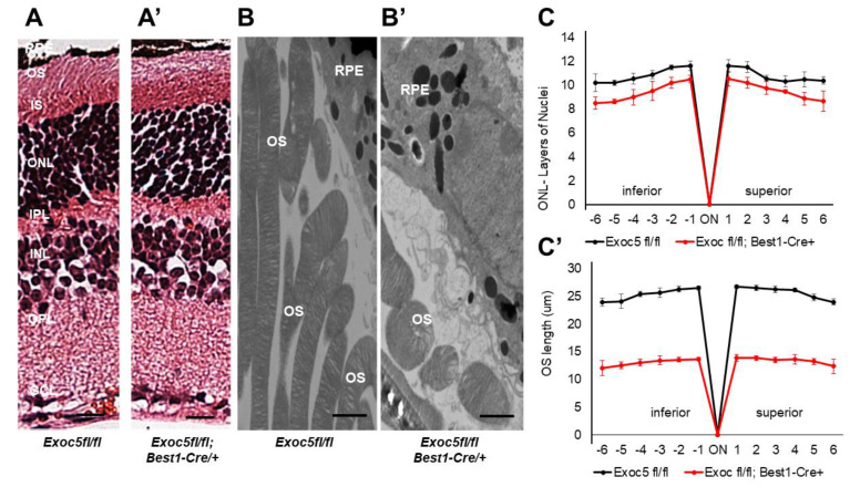
Figure 5 Histological and TEM analysis of retinas of Exoc5−/− mutants and WT mice at 27 weeks of age. (A,A’) H&E staining of WT and Exoc5−/− retinas at 27 weeks suggests the presence of disorganized rod outer segments (OS). (B,B’) Transmission electron microscopy of WT and Exoc5−/− photoreceptor cells reveal packages of rod OS in the subretinal space. (C,C’) Thickness of the ONL (C) and OS lengths (C’) from H&E sections through the optic nerve (ON; 0 μm distance from Optic Nerve and starting point) was measured at 12 locations around the retina, six each in the superior and inferior hemispheres, each equally at 150 μm distances. Scale bars = 800 nm (B,B’). OS, outer segments; RPE, retinal pigmented epithelium; IS, inner segments; ONL, outer nuclear layer; INL, inner nuclear layer; OPL, outer plexiform layer.