IMAGE

Fig. 4

ID
ZDB-IMAGE-210606-13
Source
Figures for Zhang et al., 2021
Image
Figure Caption

Fig. 4

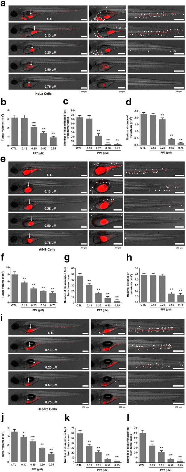
Inhibitory effects of PP7 on the invasion, dissemination and metastasis of HeLa cells, A549 cells and HepG2 cells in zebrafish xenografts. CM-Dil-labeled (red) HeLa cells (a), A549 cells (e) and HepG2 cells (i) were microinjected into the yolk sac of 2dpf embryos, and treated with different concentrations of PP7. Tumor cell invasion, dissemination and metastasis were detected using a laser scanning confocal microscope at day 3 post-injection. Arrows indicate primary tumors, white arrowheads indicate disseminated tumor foci. Scale bars represent 250 μm. Quantification of tumor volume of HeLa cells (b), A549 cells (f) and HepG2 cells (j). Quantification of numbers of disseminated tumor foci of HeLa cells (c), A549 cells (g) and HepG2 cells (k). Averages of maximal distances of metastatic foci of HeLa cells (d), A549 cells (h) and HepG2 cells (l). Data are represented as means ± SD from 3 independent experiments. n = 10/group, *P < 0.05, **P < 0.01 as compared with control

Acknowledgments
This image is the copyrighted work of the attributed author or publisher, and ZFIN has permission only to display this image to its users. Additional permissions should be obtained from the applicable author or publisher of the image. Full text @ Chin. Med.