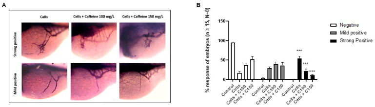
Microinjection of 3F2T cells in perivitelline space and subsequent treatment with caffeine. (A) Representative picture showing the ectopic sprouts branching out of SIVP visualized by AP assay in microinjected embryos at 72 hpf embryos (magnification 63×). (B) Percentage of embryos showing negative, mild positive and strong positive responses following microinjection of 3F2T cells, with or without caffeine treatment. In the bar diagram: CT-: negative control; Cells + C100: Cells + caffeine 100 mg/L; Cells + C150: Cells + caffeine 150 mg/L. Statistical analysis was performed with GraphPad Prism software, version 8.3.0 (GraphPad Software, Inc., La Jolla, CA, USA). *** p < 0.001, when compared to the control group.
Acknowledgments
This image is the copyrighted work of the attributed author or publisher, and
ZFIN has permission only to display this image to its users.
Additional permissions should be obtained from the applicable author or publisher of the image.
Full text @ Int. J. Mol. Sci.
Your Input Welcome
Thank you for submitting comments. Your input has been emailed to ZFIN curators who may contact you if
additional information is required.
Oops. Something went wrong. Please try again later.