IMAGE

Fig. 7

ID
ZDB-IMAGE-210527-7
Source
Figures for Mannucci et al., 2021
Image
Figure Caption

Fig. 7

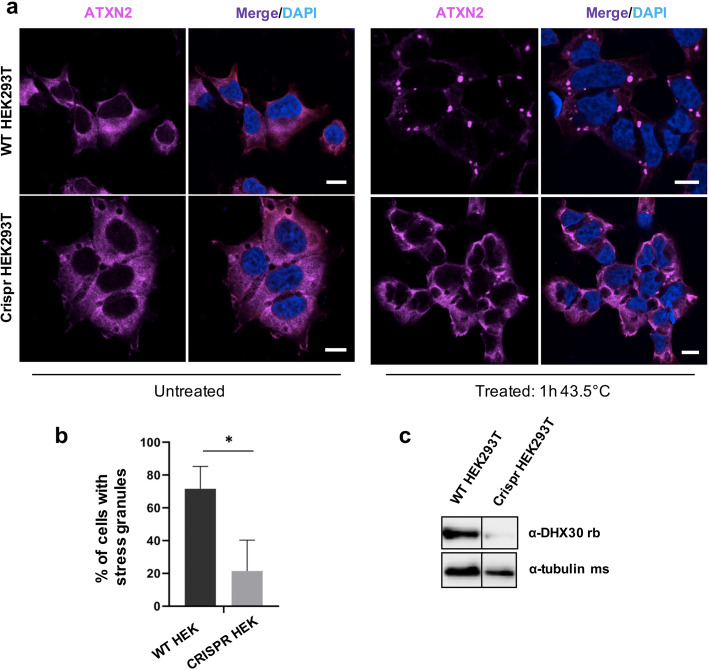
DHX30 deficiency in HEK293T cells leads to reduced formation of stress granules. a Immunocytochemical detection of endogenous ATXN2 (magenta) in WT HEK293T cells and DHX30-deficient HEK293T cells before (left panel) and after (right panel) heat shock at 43.5 °C for 1 h. Note that, upon heat stress and depletion of DHX30 (right hand, lower panel), ATXN2 does not alter its diffuse cytoplasmic distribution to accumulate in cytoplasmic foci, as observed in WT HEK293T cells (right hand, upper panel). Nuclei are identified via DAPI staining (blue). Scale bars indicate 10 μm. b Bar graph indicating the percentage of cells containing stress granules. (*: significantly different from WT HEK293T cells: *p< 0.05; n > 200 from 3 independent experiments; unpaired t test ). c Western blotting detection of DHX30 knock-out efficiency in HEK293T cells. Expression of DHX30 was reduced by 90% as detected by a DHX30 specific antibody. Tubulin was used as loading control

Acknowledgments
This image is the copyrighted work of the attributed author or publisher, and ZFIN has permission only to display this image to its users. Additional permissions should be obtained from the applicable author or publisher of the image. Full text @ Genome Med.