IMAGE

Figure 4

ID
ZDB-IMAGE-210519-34
Source
Figures for Travnickova et al., 2021
Image
Figure Caption

Figure 4

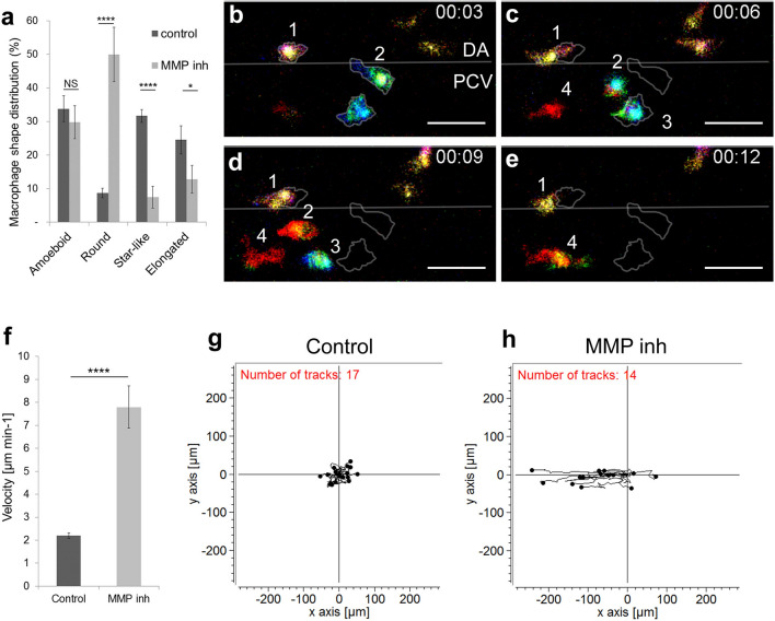
MMP-9 inhibition induces a change in macrophage shape and a transition towards an amoeboid-like migration. (a) Graph compares the macrophage shape distribution in the AGM of MMP-2 and 9 (SB-3CT) -inhibited embryos (MMP inh) to DMSO treated embryos (control) shows an increase in round shape and a decrease in star-like and elongated shapes in MMP inh embryos. N = 10 embryos for control and 15 for MMP inh. Data represent the percentage mean for each shape type out of the total number of macrophages in the AGM ± s.e.m. NS not significant; *p < 0.05; ****p < 0.0001. (be) Selected cropped images from Video 3 displays macrophage shape and migration patterns over time. Numbers point to individual macrophages, time code is expressed in hours and minutes. Grey outlines on panel C-E show the shape and position of macrophages from panel (b) (3rd minute). (f) Graph showing the velocity of macrophages in control and MMP-inhibited embryos. Data are represented as a mean ± s.e.m., n = 17 macrophages for control and 14 for MMP inhibitor from 4 different embryos, ****p < 0.0001. (g,h) Tracking plot diagram represents the migration path and distance of macrophages in the AGM in control and MMP-inhibited embryos measured every minute over 60 min. Scale in µm, n = 17 macrophages for control and 14 for MMP inhibitor from 4 different embryos. See also Video 3. Scale bar 30 µm. DA dorsal aorta, PCV posterior cardinal vein.

Acknowledgments
This image is the copyrighted work of the attributed author or publisher, and ZFIN has permission only to display this image to its users. Additional permissions should be obtained from the applicable author or publisher of the image. Full text @ Sci. Rep.