
IMAGE
FIGURE 5
- ID
- ZDB-IMAGE-210518-55
- Publication
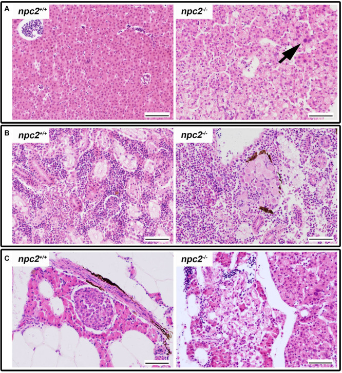
- Wiweger et al., 2021 - npc2-Deficient Zebrafish Reproduce Neurological and Inflammatory Symptoms of Niemann-Pick Type C Disease
- All Figures
- Figures for Wiweger et al., 2021
Image
|
Figure Caption
FIGURE 5
Pathological changes in soft tissues in
Figure Data
Acknowledgments
This image is the copyrighted work of the attributed author or publisher, and
ZFIN has permission only to display this image to its users.
Additional permissions should be obtained from the applicable author or publisher of the image.
Full text @ Front. Cell. Neurosci.