
|
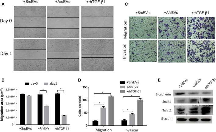
Fig. 5
The effects of sEVs on EMT activation in the recipient cells. (A) After cell‐sEV incubation, the ability of wound closure of the recipient cells was measured using cell wound‐healing assay. (B) 24 h after incubation, unclosed areas were calculated. (C) After cell‐sEV incubation, cell invasion and migration capacities were analyzed by transwell assay. (D) The numbers of invaded and migrated cells were plotted. (E) The EMT markers expressed in the recipient cells were measured by western blots. Treatment of rhTGF‐β1 was used as a positive control. All experiments were repeated, and more than three replicates were included in A–D. The data were presented as the mean ± standard deviation. *