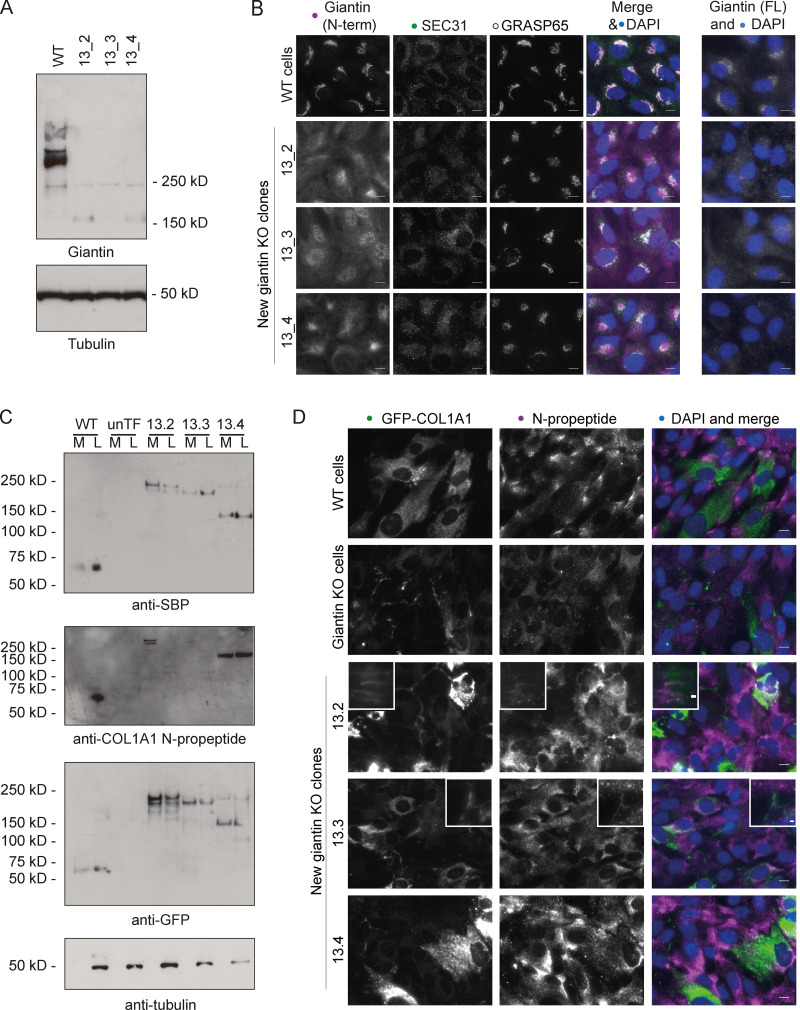
Validation of processing defects in other giantin mutant RPE1 lines. (A) Immunoblot of lystates from WT and giantin exon 13 KO RPE1 clones probed for giantin (polyclonal against full-length protein) and tubulin (housekeeping). (B) Widefield maximum-projection images of WT and giantin KO RPE1 cell clones immunolabeled for the N-terminus (N-term) of, and full-length (FL) giantin; ER exit sites (Sec31); and the Golgi (GRASP65) as indicated. Scale bar = 10 µm. (C) Immunoblots of secretion assays performed on WT and giantin KO clones stably expressing GFP-COL1A1 and untransfected giantin KO cells (unTF). Blots show the medium (M) and lysate (L) fractions of each cell population immunoblotted for GFP, SBP, and pro-α1(I) N-propeptide as indicated. (D) Maximum-projection widefield images of GFP signal in giantin KO clones stably expressing GFP-COL1A1 and immunolabeled for pro-α1(I) N-propeptide (magenta). Scale bar = 10 µm.
Acknowledgments
This image is the copyrighted work of the attributed author or publisher, and
ZFIN has permission only to display this image to its users.
Additional permissions should be obtained from the applicable author or publisher of the image.
Full text @ J. Cell Biol.
Your Input Welcome
Thank you for submitting comments. Your input has been emailed to ZFIN curators who may contact you if
additional information is required.
Oops. Something went wrong. Please try again later.