IMAGE

Figure 5

ID
ZDB-IMAGE-210508-14
Source
Figures for DeLaurier et al., 2021
Image
Figure Caption

Figure 5 Embryonic disintegration in <italic>ctnnd1</italic> mutants.

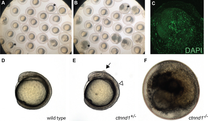
(A, B) Frames from time-lapse imaging (Movie S1) illustrate disintegrating phenotype of ctnnd1 mutants. Asterisk(s) mark two of the embryos that disintegrated during the time-lapse recording. (C) DAPI staining of fixed ctnnd1 mutant embryos reveals that cells with intact nuclei dissociate from the embryo. (D–F) Phenotypes of ctnnd1 sibling embryos at the 6-somite stage. The homozygous mutant (F) has disintegrated, the heterozygous mutant (E) displays clumps of cells along the dorsal surface, and the wild-type embryo appears normal (D). The solid arrow marks a clump of cells dorsal to the midbrain and the open arrowhead marks a clump of cells dorsal to the hindbrain in the heterozygote.

Acknowledgments
This image is the copyrighted work of the attributed author or publisher, and ZFIN has permission only to display this image to its users. Additional permissions should be obtained from the applicable author or publisher of the image. Full text @ Peer J.