Image
Figure Caption
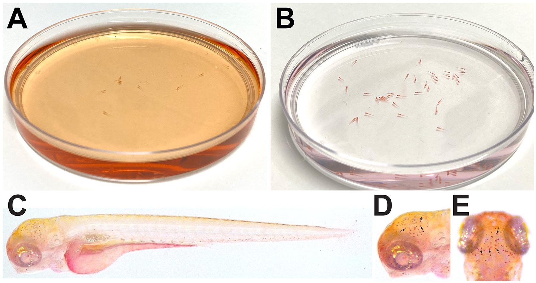
Fig. 3 A. Color of fish water after addition of neutral red. B. Color of fish and water after removal of neutral red. C. Full body image of control uninjected 4 dpf larvae after neutral red staining. D and E. Lateral (D) and dorsal (E) view of neutral red staining. C-E. Larvae imaged in 3% methyl cellulose. Black arrows point to individual microglial cells.
Acknowledgments
This image is the copyrighted work of the attributed author or publisher, and
ZFIN has permission only to display this image to its users.
Additional permissions should be obtained from the applicable author or publisher of the image.
Full text @ Bio Protoc

