
Image
|
Figure Caption
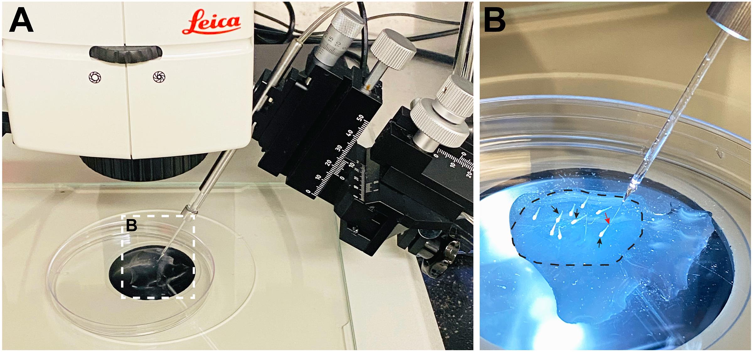
Fig. 2 A. Mounted larvae are placed under a fluorescent stereomicroscope adjacent to a microinjection needle attached to a micro-manipulator. B. Higher magnification of mounted larvae. Dotted circle shows solidified thin layer of low-melt agarose covered with a small pool of system water supplemented with PTU. Black arrows point to larvae. Red arrow points to tip of needle (Note: This arrow is not pointing to the injection site.)
Acknowledgments
This image is the copyrighted work of the attributed author or publisher, and
ZFIN has permission only to display this image to its users.
Additional permissions should be obtained from the applicable author or publisher of the image.
Full text @ Bio Protoc