
|
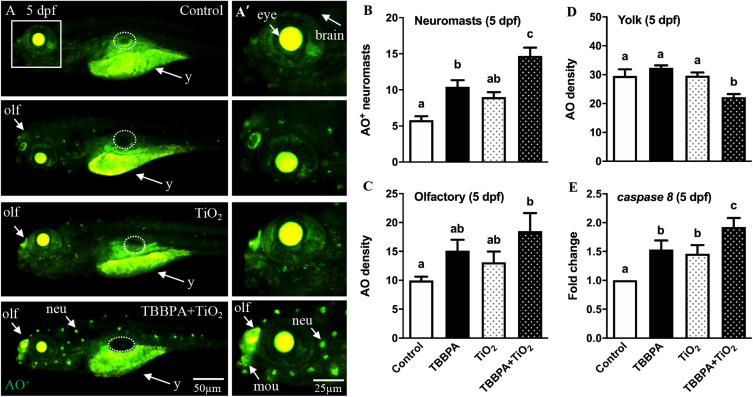
Fig. 5 Effect of TBBPA and TiO2 NP on cell apoptosis. Zebrafish embryos were exposed to 0.1% DMSO control, 2 μM TBBPA, 0.1 mg/L TiO2 NP and TBPPA/TiO2 NP mixture from 8 to 120 hpf. Larvae were harvested immediately after exposure and used for cell apoptosis analysis. (A) Representative images showing acridine orange (AO) staining for larvae at 5 dpf. (A’) Images of the head region (within the white square in A) at a higher magnification. (B) The number of AO positive neuromasts in the lateral line system of 5 dpf larvae. (C-D) Quantification of AO staining density in the olfactory (C) and yolk region (D) (n = 20). (E) Gene expression fold changes of cell apoptotic marker caspase 8 (casp8) at 120 hpf (n = 3, each replicate consists pooled RNA sample from 20 larvae). The dashed elliptical area marked the swim bladder region. olf: olfactory epithelium; neu: neuromast; mou: mouth; y: yolk region. Values plotted are mean ± SEM and bars sharing the same letter indicate no significant difference at the level of P < 0.05 using one-way analysis of variance (ANOVA) followed by Tukey’s multiple comparison test.