
|
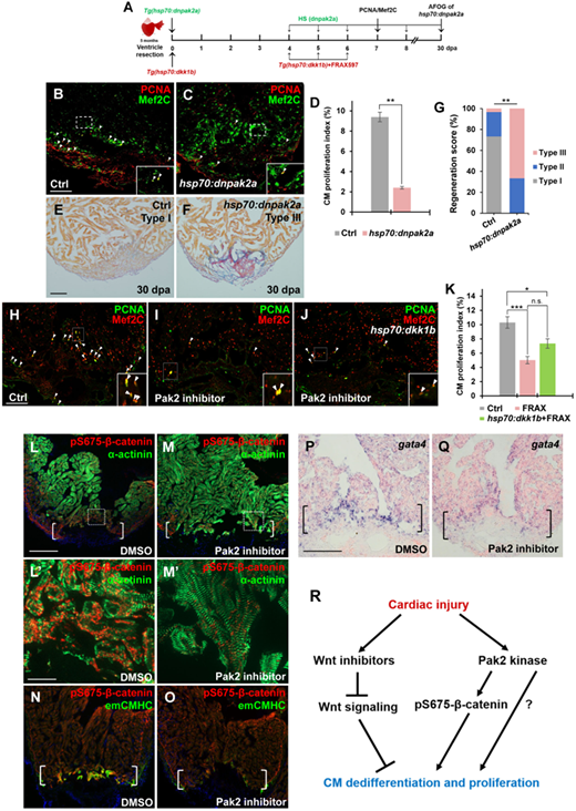
Fig. 7 Inhibition of Pak2 activity impairs CM dedifferentiation and proliferation. (A) Schematic of heat shock experiments and FRAX597 treatment for PCNA/Mef2C analyses. Heat shock: 37°C for 2 h daily from 4 to 6 dpa for PCNA/Mef2C assay or over the time period from 4 to 30 dpa for AFOG analysis; treatment: 1 µM FRAX597 or 0.1% DMSO from 4 to 6 dpa. (B‒D) Confocal image analyses displaying PCNA+Mef2C+ cells (arrowheads, B and C) and PCNA-labeled CM proliferation index (D) in Ctrl and Tg(hsp:dnpak2a) hearts at 7 dpa. Insets indicate high-magnification images in dash-lined areas. Data are mean ± SEM from five hearts for each group. Student’s t-test, **P < 0.01. (E‒G) Representative images (E and F) and quantification of regeneration scores (G) of ventricle sections from heat-shocked Ctrl and Tg(hsp70:dnpak2a) animals at 30 dpa and stained with AFOG. Orange: muscle; blue: collagen; red: fibrin. Fisher’s exact test. **P < 0.01. (H‒K) Confocal image analyses displaying PCNA+Mef2C+ cells (arrowheads) and quantification of CM proliferation indices (K) in DMSO-treated (H, Ctrl), Pak2 inhibitor (FRAX597)-treated (I), and FRAX597-treated Tg(hsp:dkk1b) (J) hearts at 7 dpa. Insets indicate high-magnification images of dash-lined area. Data are represented as mean ± SEM from five hearts for each group. Statistical significance was calculated using one-way ANOVA followed by Tukey’s test. n.s., none significance; *P < 0.05, ***P < 0.001. (L‒O) Immunostaining analyses displaying reduction of pS675-β-catenin (L and M) and emCMHC (N and O) at apical myocardial cells of the wounded edge in FRAX597-treated hearts compared to DMSO-treated hearts at 7 dpa. (L′ and M′) High-magnification images of dash-lined windows in L and M, respectively, exhibiting disassembled sarcomeres in DMSO-treated hearts (L′) and relatively normal striated sarcomeres in FRAX597-treated hearts (M′). (P and Q) ISH analysis displaying a reduction of gata4 at the wounded myocardial cell edge in FRAX597-treated hearts compared to DMSO-treated hearts at 7 dpa. Brackets indicate amputation planes. Scale bar, 100 µm (except 10 µm in L′ and M′). (R) A working model for Wnt/Pak2/pS675-β-catenin signaling events regulating heart regeneration. Cardiac injury induces multiple Wnt antagonists that restrain Wnt signaling throughout the wounded heart, enabling CM dedifferentiation and proliferation. In the context of Wnt signaling inhibition, Pak2 is upregulated in cardiac wounds, where it phosphorylates cytoplasmic β-catenin at the Ser 675 residue and increases its stability in disassembled sarcomeres, enhancing CM renewal and heart regeneration.