IMAGE

Fig 4

ID
ZDB-IMAGE-210503-149
Source
Figures for Wright et al., 2021
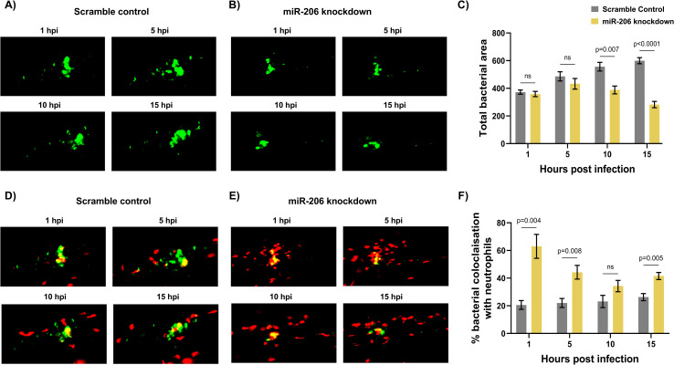
Image
Figure Caption

Fig 4 Increased miR-206 knockdown-associated neutrophils prevent bacterial dissemination.

(A-B) Representative images of bacterial granulomas in trunk-infected control and miR-206 knockdown embryos. (C) Quantification of M. marinum burden in trunk-infected control and miR-206 knockdown embryos. (D-E) Representative images of M. marinum-neutrophil interactions in trunk-infected control and miR-206 knockdown embryos. Neutrophils are red Tg(lyzC:dsred) and M. marinum is green (wasabi); co-localisation is indicated by yellow fluorescence. (F) Quantification of the proportion of the bacterial fluorescence overlapping with neutrophil fluorescence (co-localisation yellow fluorescence in D-E) in trunk-infected control and miR-206 knockdown embryos. Each data point represents the mean of 6 foci of infection from 6 separate embryos with SEM shown. Differences between groups was calculated using multiple t-tests and the Holm-Sidak method.

Acknowledgments
This image is the copyrighted work of the attributed author or publisher, and ZFIN has permission only to display this image to its users. Additional permissions should be obtained from the applicable author or publisher of the image. Full text @ PLoS Pathog.