
|
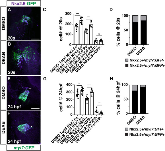
Fig. 4 RA restricts the size of the FHF and maintains the SHF progenitor population in DEAB-treated embryos. (A, B) IHC for Nkx2.5 (purple) and GFP (myl7:GFP - green) in DMSO- and DEAB-treated embryos at the 20s stage (19 hpf). The cardiac cone was sometimes delayed in forming in DEAB-treated embryos relative to DMSO-treated control embryos. Views are dorsal with anterior up. Scale bar – 100 μm. (C) Quantification of the number of Nkx2.5+/GFP+ and Nkx2.5+/GFP- cells in DMSO- and DEAB-treated embryos at the 20s stage (19 hpf). ∗∗∗ indicates p ≤ 0.0004. (D) Percentage of Nkx2.5+/GFP+ and Nkx2.5+/GFP- cells within the hearts of DMSO- and DEAB-treated embryos at the 20s stage (19 hpf). (E, F) IHC for Nkx2.5 (purple) and GFP (myl7:GFP - green) in DMSO- and DEAB-treated embryos at 24 hpf. Frontal views with the arterial pole up. Arrows indicate the border between Nkx2.5+/GFP+ and Nkx2.5+/GFP- cells. Scale bar – 100 μm. (G) Quantification of the number of Nkx2.5+/GFP+ and Nkx2.5+/GFP- cells in DMSO- and DEAB-treated embryos at 24 hpf. ∗∗ indicates p ≤ 0.0060, ∗∗∗∗ indicates p < 0.0001. (H) Percentage of Nkx2.5+/GFP+ and Nkx2.5+/GFP- cells within the hearts of DMSO- and DEAB-treated embryos at 24 hpf.
Reprinted from Developmental Biology, 473, Duong, T.B., Holowiecki, A., Waxman, J.S., Retinoic acid signaling restricts the size of the first heart field within the anterior lateral plate mesoderm, 119-129, Copyright (2021) with permission from Elsevier. Full text @ Dev. Biol.