Image
Figure Caption

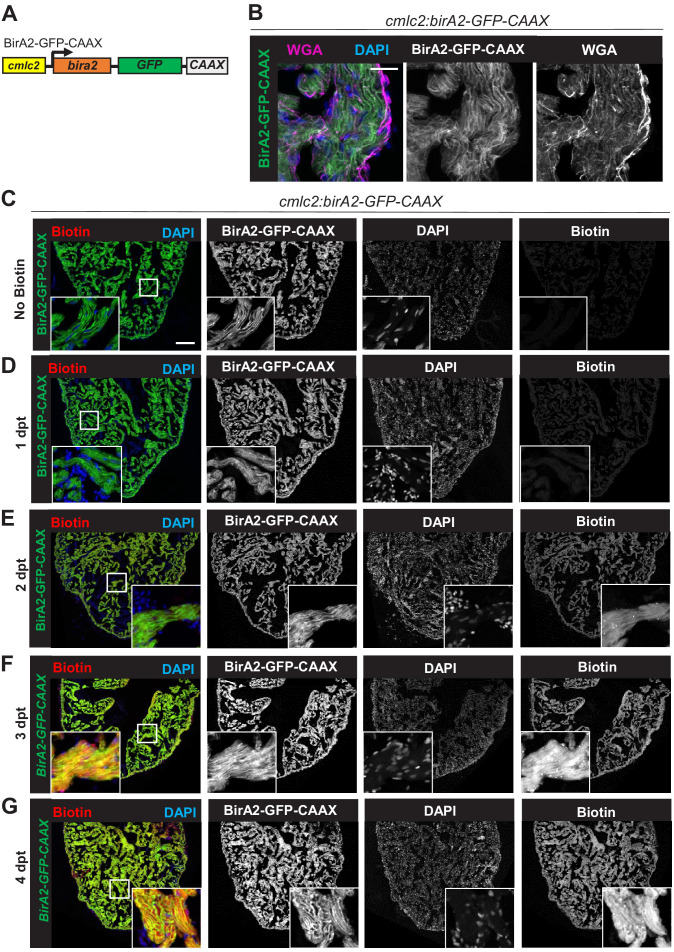
Figure 1—figure supplement 2. Evaluation of BirA2-GFP-CAAX localization and biotinylation.

(A) BirA2-GFP-CAAX is expressed in cardiomyocytes via the cmlc2 promoter. (B) BirA2-GFP-CAAX is localized to the cell membrane. Membrane staining with wheat germ agglutinin showed colocalization with the BirA2-GFP-CAAX signal. Scale bar: 10 µm. (C–G) Immunofluorescence images of sections of ventricles with native BirA2-GFP-CAAX signal stained for DAPI. Animals were injected intraperitoneally with biotin for 1, 2, 3, or 4 days. dpt: days post treatment. Scale bar: 50 µm.

Acknowledgments
This image is the copyrighted work of the attributed author or publisher, and ZFIN has permission only to display this image to its users. Additional permissions should be obtained from the applicable author or publisher of the image. Full text @ Elife