
|
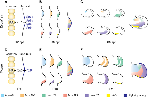
Fig. 3 Early patterning of limb and pectoral fin buds is comparable. (A–C) Early zebrafish fin bud patterning. (A) Fin induction at 12 hpf: RA from the somites activates signals that lead to activation of tbx5 in the lateral plate and Fgf signaling in the distal bud, establishing the AER. (B) Early expression of Hox genes and shh at 30 hpf. (C) Expression of Hox genes and shh at 60 hpf. Note that the hoxd11 expression domain in the posterior fin bud region remains restricted to this region at 30 hpf and 60 hpf. Dashed line = boundary of fin bud proper and fin fold. (D–F) Early mouse limb bud patterning. (D) Similar induction signals as described in A for the fin bud occur in the mouse at E9. (E) Early expression of Hox genes and shh in E10.5 mouse limbs shows a similar pattern observed as in B for the fin bud. While hoxd9 and hoxd10 extend through the whole bud, hoxd11, hoxd12, hoxd13, and shh are restricted to the posterior domain. (F) Hox expression domain in E12.5 mouse limbs differs from those observed in C in the fin bud; hoxd11, hoxd12, and hoxd13 domains extend more anteriorly; and shh remains restricted to the posterior region. Left to right = proximodistal axis, top down = anterior–posterior axis. E10.5 = embryonic day 10.5; E12.5 = embryonic day 12.5; E9 = embryonic day 9; hpf = hours post fertilization.