
|
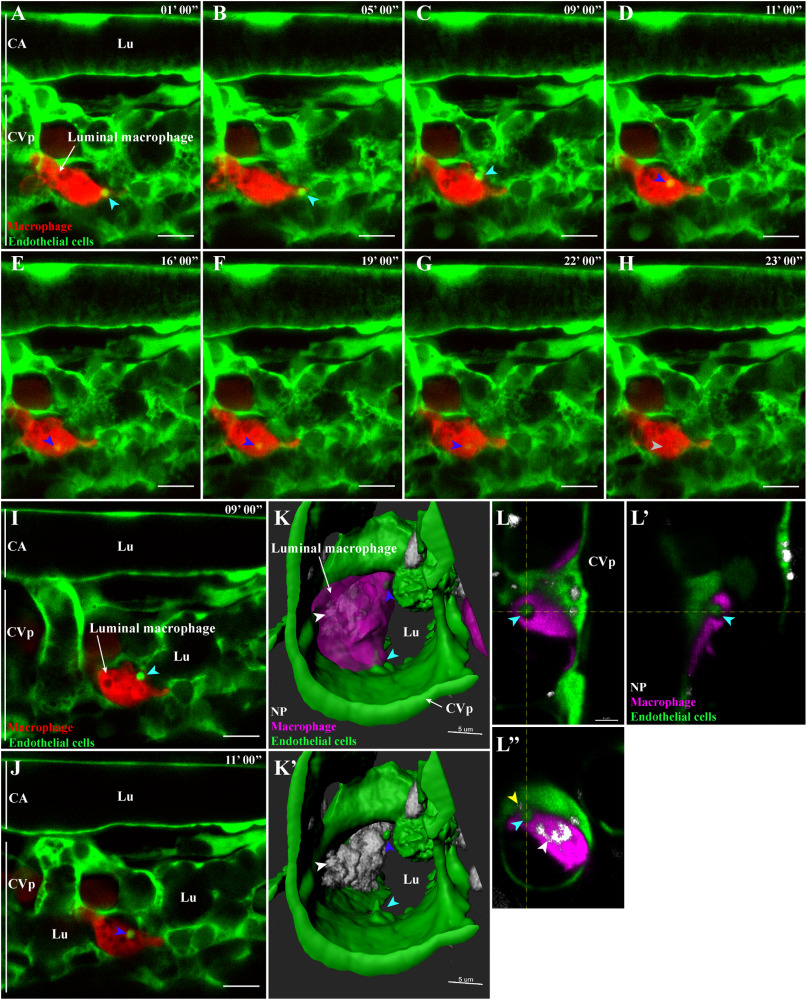
Fig. 6 Microvesicle-like transfer from endothelial cells to macrophages. Representative live-acquisitions of 3 dpf mpeg1:mCherry and fli:GFP double transgenic zebrafish, 30 min after the intravenous injection of far-red fluorescent PLA-NP and using a spinning-disk confocal microscope. The acquisitions have been made in the tail region, at the caudal vein plexus. Endothelial cells, macrophages and PLA-NP are highlighted in green, red and white, respectively. (A-B) At the start of the video (9 μm MIP), close to a macrophage residing in the bloodstream, an endothelial cell from the caudal vein plexus is forming a bright GFP+ microvesicle-like structure (cyan arrowhead). (C) Nine minutes later, the microvesicle-like structure is released from the endothelial cell (cyan arrowhead), it is then quickly internalized by the adjacent macrophage (D - blue arrowhead). Once internalized, the fluorescence of the microvesicle-like structure rapidly fades (E-G - blue arrowheads) to finally disappear 13 min after its internalization (H – grey arrowhead). Optical sections (1 μm) from (C) and (D), (I) highlights the contact between the GFP+ microvesicle and the plasma membrane of the macrophage (cyan arrowhead), while (J) emphasize its internalization inside a mCherry negative cellular compartment (blue arrowhead). (K-K′) A luminal macrophage, positive for PLA-NP (white arrowhead) and with a GFP+ cellular compartment (blue arrowhead) is wrapping around a budding microvesicle-like structure of an endothelial cell from the caudal vein plexus (cyan arrowhead). Fluorescence are represented as 3D reconstructions. (L-L") Orthogonal views from (K) reveal the presence of PLA-NP in close proximity to the budding-microvesicle-like structure (yellow arrowhead). Annotations: CA, caudal artery; CVp, caudal vein plexus and Lu, lumen. Scale bars: 10 um (A-J), 5 μm (K-K′) and 4 μm (L). (For interpretation of the references to colour in this figure legend, the reader is referred to the web version of this article.)