
FIGURE 1
- ID
- ZDB-IMAGE-210403-8
- Publication
- Moreau et al., 2021 - Deciphering DSC2 arrhythmogenic cardiomyopathy electrical instability: From ion channels to ECG and tailored drug therapy
- All Figures
- Figures for Moreau et al., 2021
|
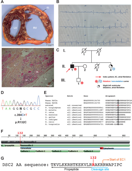
FIGURE 1
Clinical presentation. (A) Upper panel: transverse section of the ACM patient's explanted heart. The right ventricle (RV) is dilated. Adipo–fibrotic replacement of the myocardium is also observed. Lower panel: Microscopic section of the patient's explanted heart stained with hematoxylin, eosin and safran. Inflammation, adipose tissue and myocardial necrosis are observed. IT, interstitial tissue; INF, inflammation; FI, adipose tissue; MN, myocardial necrosis. (B) Twelve‐lead surface ECG illustrating the patient's electrical profile. The patient was in sinus rhythm with a PR interval at 160 ms, QRS interval at 120 ms and S wave slurring. Negative T waves were observed in V2 through V5. (C) Family pedigree. The index patient (II.1) is indicated by a red arrow. Individuals indicated with a red cross carry the mutation (II‐1 and III‐1). (D) Electropherogram of the patient's DNA showing the