
Fig. 1
- ID
- ZDB-IMAGE-210402-7
- Publication
- Square et al., 2021 - Distinct tooth regeneration systems deploy a conserved battery of genes
- All Figures
- Figures for Square et al., 2021
|
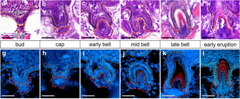
Fig. 1
Stickleback pharyngeal tooth morphogenesis. All images show sagittal or transverse sections from sticklebacks (20–40 mm standard length). The basalmost layer of epithelium is outlined with yellow dashed lines in all panels, the mesenchyme is outlined in red. During late bell stages (