IMAGE

FIGURE 1

ID
ZDB-IMAGE-210319-93
Source
Figures for Cheung et al., 2021
Image
Figure Caption

FIGURE 1

Phalloidin staining reveals the presence of actin-rich rod-like projections, distinct from OSN microvilli and cilia, in the zebrafish larval and juvenile olfactory epithelium.
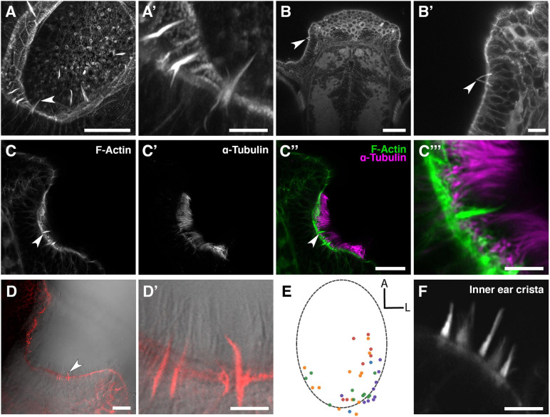
 (A) Maximum intensity projection of an Airyscan confocal image of phalloidin stain in an olfactory pit of a 5 dpf wild-type larva; anterior to the top right, lateral to the bottom right. Arrowhead marks one example olfactory rod. Scale bar = 20 μm. (A′) Enlargement of olfactory rods in panel (A). Scale bar = 5 μm. (B) Dorsal view low power image of phalloidin stain in the head of an 18 dpf (5 mm) wild-type juvenile zebrafish; anterior to the top. Arrowhead marks the position of two olfactory rods in an olfactory pit. Scale bar = 50 μm. (B′) Enlargement of OE in panel (B). Arrowhead marks two olfactory rods. Scale bar = 10 μm. (C–C″) Airyscan confocal image of Alexa-phalloidin signal (C), acetylated α-tubulin immunohistochemistry signal (C′), and merged signals (C″) in an olfactory pit of a 4 dpf wild-type larva; anterior to the top, lateral to the right. Arrowhead marks one example olfactory rod. Scale bar = 20 μm. (C″′) Enlargement of olfactory rod in panel (C″). Scale bar = 5 μm. (D) Differential interference contrast (DIC) image and phalloidin stain (red) in an olfactory pit of a 5 dpf wild-type larva; anterior to the top, lateral to the right. Arrowhead marks one example olfactory rod. Scale bar = 20 μm. (D′) Enlargement of olfactory rods in panel (D). Surrounding olfactory cilia are visible and unlabelled by Alexa-phalloidin. Scale bar = 5 μm. (E) A map of the positions of olfactory rod cell projection bases in olfactory pits of 4 dpf wild-type larvae (N of olfactory pits = 5), based on 2D maximum intensity projections of confocal images of phalloidin stains; anterior “A” to the top, lateral “L” to the right. One dot represents one olfactory rod. Different coloured dots represent olfactory rods from different larvae. (F) Airyscan confocal image of phalloidin stain in an inner ear crista of a 5 dpf wild-type larva. Hair cell stereocilia are labelled with Alexa-phalloidin, and are arranged in a stepped array. In the stereociliary bundle on the extreme left, four different stereociliary lengths are visible [compare with panel (A′)]. Scale bar = 5 μm.

Acknowledgments
This image is the copyrighted work of the attributed author or publisher, and ZFIN has permission only to display this image to its users. Additional permissions should be obtained from the applicable author or publisher of the image. Full text @ Front. Physiol.