
Fig. 6
- ID
- ZDB-IMAGE-210316-12
- Publication
- Bertozzi et al., 2020 - Is zebrafish heart regeneration "complete"? Lineage-restricted cardiomyocytes proliferate to pre-injury numbers but some fail to differentiate in fibrotic hearts
- All Figures
- Figures for Bertozzi et al., 2020
|
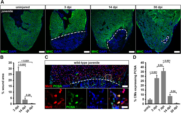
Fig. 6 Fig. 6. Juvenile hearts regenerate after cryoinjury via cardiomyocyte proliferation. (A) Myosin heavy chain (MHC) immunostaining reveals wound area (dashed lines) on heart sections of cryoinjured juvenile fish. Scale bars, 100 μm. (B) Wound area (MHC— area) relative to the whole ventricle area is plotted. Error bars represent s.e.m. n (hearts) = 6 for all time-points. One-way ANOVA + Holm-Sidak’s multiple comparisons test. (C) PCNA+ Mef2+ cardiomyocytes in juvenile hearts at 7 dpi. Scale bars, 50 μm (overview), 12.5 μm (magnified view). (D) The average percentage of cardiomyocytes expressing PCNA within 150 μm from the wound border is plotted. Error bars represent s.e.m. n (hearts) = 6 for all time-points. One-way ANOVA + Holm-Sidak’s multiple comparisons test.
Reprinted from Developmental Biology, 471, Bertozzi, A., Wu, C.C., Nguyen, P.D., Vasudevarao, M.D., Mulaw, M.A., Koopman, C.D., de Boer, T.P., Bakkers, J., Weidinger, G., Is zebrafish heart regeneration "complete"? Lineage-restricted cardiomyocytes proliferate to pre-injury numbers but some fail to differentiate in fibrotic hearts, 106-118, Copyright (2020) with permission from Elsevier. Full text @ Dev. Biol.