
Fig. 4
- ID
- ZDB-IMAGE-210316-10
- Publication
- Bertozzi et al., 2020 - Is zebrafish heart regeneration "complete"? Lineage-restricted cardiomyocytes proliferate to pre-injury numbers but some fail to differentiate in fibrotic hearts
- All Figures
- Figures for Bertozzi et al., 2020
|
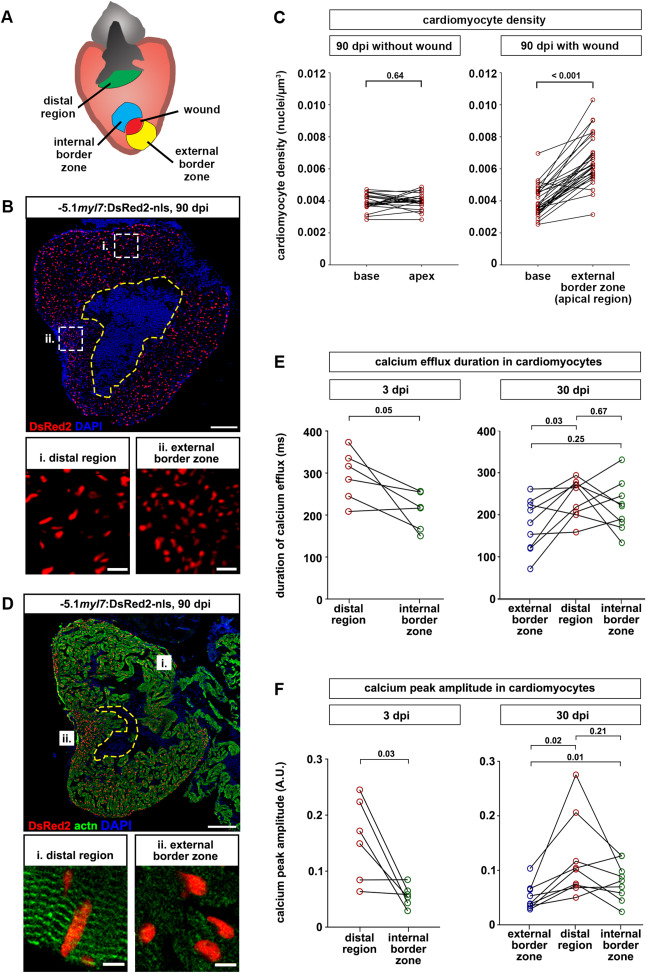
Fig. 4 Fig. 4. Cardiomyocytes are denser and less mature at the external wound border zone in scarred hearts. (A) Cartoon depicting different regions of ventricles containing residual scars that were analyzed. (B) Cardiomyocyte nuclei density is higher in the external wound border zone (box ii) than the distal region (box i) at 90 dpi. The wound is outlined with a dashed yellow line. Scale bar, 100 μm. (C) At 90 dpi, cardiomyocyte density at the base and apex does not differ in hearts without wound, but is higher at the external wound border in hearts with wounds. n (hearts) = 22 (without wound), 30 (wound). Paired two-tailed t-test. (D) Sarcomeric structures cannot be identified by alpha-actinin staining in the external wound border in hearts with residual scars. n (hearts with assembled sarcomeres): wound border, 2/13 hearts at 30 dpi, 4/23 hearts at 90 dpi; distal myocardium, 12/13 hearts at 30 dpi, 21/23 hearts at 90 dpi. The wound is outlined with a dashed yellow line. Scale bar, 100 μm. (E, F) Calcium efflux duration (E) and amplitude of calcium peaks (F) in myl7:GCaMP6f-nls-T2A-RCaMP107-NES-pA)hu11799 cardiomyocytes is plotted at 3 dpi and at 30 dpi for hearts containing residual scars. n (hearts) = 6 (3 dpi), 9 (30 dpi). Paired two-tailed t-test.
Reprinted from Developmental Biology, 471, Bertozzi, A., Wu, C.C., Nguyen, P.D., Vasudevarao, M.D., Mulaw, M.A., Koopman, C.D., de Boer, T.P., Bakkers, J., Weidinger, G., Is zebrafish heart regeneration "complete"? Lineage-restricted cardiomyocytes proliferate to pre-injury numbers but some fail to differentiate in fibrotic hearts, 106-118, Copyright (2020) with permission from Elsevier. Full text @ Dev. Biol.