Figure Caption
Fig. 3
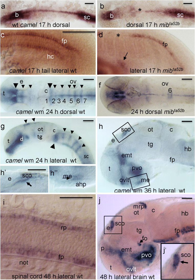
The expression pattern of camel in wild-type embryos and mutants detected by whole-mount in situ hybridization during neurogenesis. camel is expressed in the axial structures (fp, rp, hp), along segmental boundaries in the brain and in circumventricular organs. a, c 17 hpf, wild-type embryos. b, d 17 hpf, mibta52b mutant. e, g 24 hpf, wild-type embryos. f 24 hpf, mibta52b mutant. h 36 hpf, brain of a wild-type embryo. i Wild-type trunk, 48 hpf. h, j Wild-type brain, 48 hpf. Numbers define rhombomeres of the hindbrain, asterisk indicates gaps in the floor plate, and arrow indicates remains of hypochord. a, b, e, f Dorsal view. c, d, g–j Lateral view. Abbreviations: ahp, adenohypophysis; b, brain; c, cerebellum; d, diencephalon; e, epiphysis; emp, eminentia thalami; epIII, ependyma of the third ventricle; ey, eye; hb, hindbrain; ht, hypothalamus; fo, flexural organ; fp, floor plate; hb, hindbrain; hc, hypochord; ht, hypothalamus; me, median eminence; mrp, midbrain roof plate; not, notochord; ot, optic tectum; ov, otic vesicle; ovlt, organum vasculosum lamina terminalis; pvo, paraventricular organ; rp, roof plate; sc, spinal cord; sco, subcommissural organ; t, telencephalon; tg, tegmentum. Scale bar = 100 μm
Acknowledgments
This image is the copyrighted work of the attributed author or publisher, and
ZFIN has permission only to display this image to its users.
Additional permissions should be obtained from the applicable author or publisher of the image.
Full text @ Cell Tissue Res.