
|
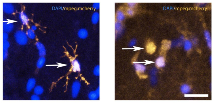
Figure 4 Resting and activated microglia under injured and uninjured (control) conditions in the telencephalon of zebrafish. Confocal microscopy showing quiescent (resting) microglia (left panel) and ameboid (activated) microglia (right panel) in the adult zebrafish telencephalon. There is an obvious change in the shape of the microglia between injured and uninjured tissue, illustrated by the mpeg:mcherry transgenic fish line, which labels microglia in the central nervous system. Arrows show the resting morphology of microglia cells (left panel) the ameboid shape of activated microglia at 1dpl (right panel). Bar: 18 μm.