IMAGE

Fig. 8

ID
ZDB-IMAGE-210307-31
Source
Figures for Ni et al., 2021
Image
Figure Caption

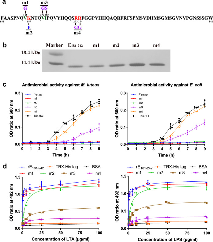
Fig. 8 Antibacterial activity of rE<sub>181–242</sub> mutants.

a Diagram showing the site-directed mutagenesis. b SDS-PAGE of the mutants, showing that they all have a similar molecular mass. c Antibacterial activities of rE181–242 mutants against M. luteus and E. coli. rE181–242 was used as positive control and Tris-HCl buffer used as negative control. d Interaction of E1–180 mutants with LTA and LPS revealed by ELISA. Each point in the graph represents the mean ± SD (n = 3). The data are from three independent experiments performed in triplicate. The bars represent the mean ± SD.

Acknowledgments
This image is the copyrighted work of the attributed author or publisher, and ZFIN has permission only to display this image to its users. Additional permissions should be obtained from the applicable author or publisher of the image. Full text @ Commun Biol