IMAGE

Fig. 3

ID
ZDB-IMAGE-210307-26
Source
Figures for Ni et al., 2021
Image
Figure Caption

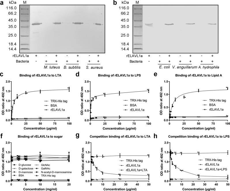
Fig. 3 Binding of rELAVL1a to Gram-positive and Gram-negative bacteria, LTA, LPS, and lipid A.

a, b Western blotting about interaction of rELAVL1a with Gram-positive (a) and Gram-negative (b) bacteria. Lane M, marker; lane 1, purified rELAVL1a; lane 2, 4 and 6, M. luteus, B. subtilis and S. aureus (a) or E. coli, V. anguillarum and A. hydrophila (b) incubated in the presence of rELAVL1a; lane 3, 5, and 7, M. luteus, B. subtilis and S. aureus (a) or E. coli, V. anguillarum and A. hydrophila (b) incubated in the absence of rELAVL1a. c, d, e Interaction of rELAVL1a with LTA (c), LPS (d), Lipid A (e) revealed by ELISA. f Effects of various sugars on the interaction of rELAVL1a with LPS. g, h Interaction of rELAVL1a with LPS was inhibited by LTA (g) and LPS (h), the final concentration of rELAVL1a was 25 μg/ml. Each point in the graph represents the mean ± S.D. (n = 3). The data are from three independent experiments performed in triplicate. The bars represent the mean ± S.D.

Acknowledgments
This image is the copyrighted work of the attributed author or publisher, and ZFIN has permission only to display this image to its users. Additional permissions should be obtained from the applicable author or publisher of the image. Full text @ Commun Biol