
Image
|
Figure Caption
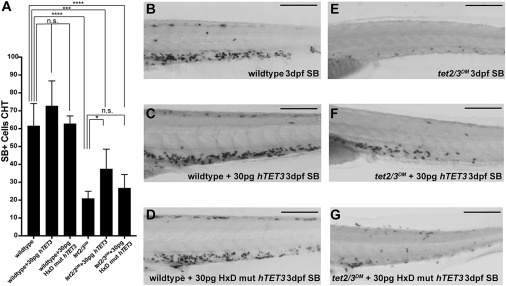
Fig. 2 Figure 2. Human TET3 Overexpression Rescues the Granulation Defect in tet2/3DM Embryos (A) Quantification of SB+ cells in the CHT region for uninjected, 30 pg wild-type hTET3 and 30 pg catalytically dead mutant (HxD) hTET3-injected wild-type and tet2/3DM embryos at 3 dpf; n = 6/condition, ∗p < 0.05, ∗∗∗p < 0.001, ∗∗∗∗p < 0.0001 by 2-way ANOVA with Tukey post hoc test. (B–G) Representative images of the CHT of SB-stained uninjected, wild-type hTET3-injected, or mutant hTET3-injected wild-type and tet2/3DM embryos at 3 dpf. Scale bars represent 200 μm.
Acknowledgments
This image is the copyrighted work of the attributed author or publisher, and
ZFIN has permission only to display this image to its users.
Additional permissions should be obtained from the applicable author or publisher of the image.
Full text @ Cell Rep.