
|
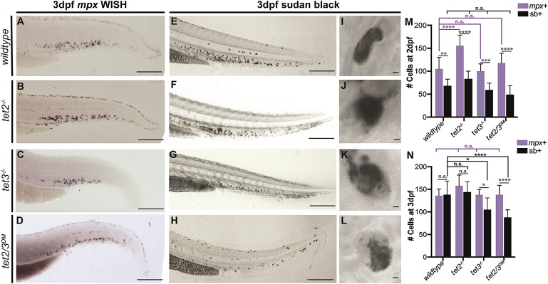
Fig. 1 Figure 1. Granulation Is Defective in tet3−/− and tet2/3DM Embryonic Neutrophils Representative images of neutrophil staining in wild-type, tet2−/−, tet3−/−, and tet2/3DM embryos at 3 dpf. (A–D) Caudal hematopoietic tissue (CHT) region following WISH to analyze mpx transcripts. (E–H) CHT region of embryos stained for Sudan black (SB). Scale bars in (A)–(H) represent 200 μm. (I–L) High-resolution images of SB-stained neutrophils. Scale bars represent 2 μm. (M and N) Quantification of total mpx+ (immature and mature neutrophils) and SB+ (mature neutrophils) cells in embryos at 2 dpf (M) and 3 dpf (N); n = 10/condition, ∗p < 0.05, ∗∗∗p < 0.001, ∗∗∗∗p < 0.0001 by 2-way ANOVA with Tukey post hoc test.