
|
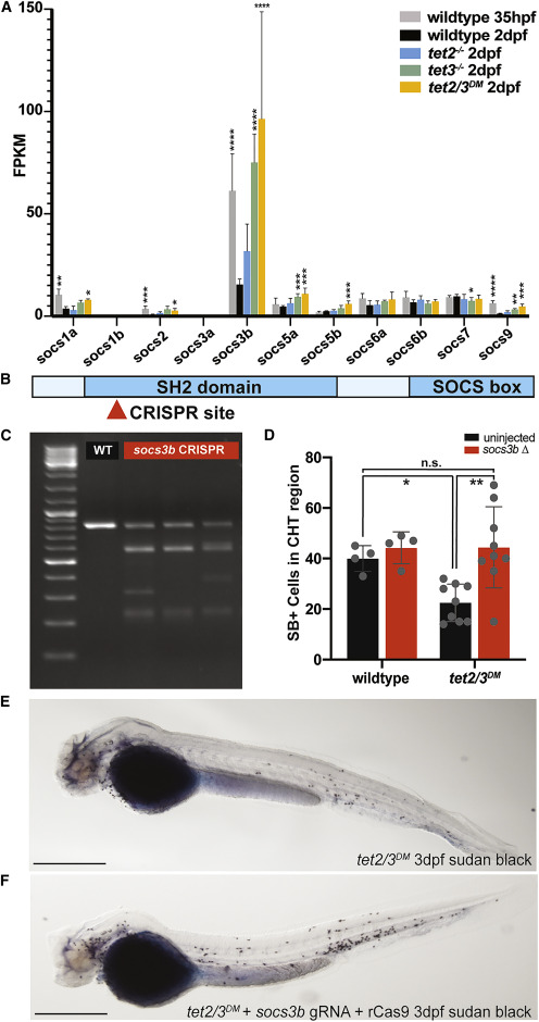
Fig. 6 Figure 6. Socs3b mRNA Levels Are Increased in tet Mutant Neutrophils and Mutation of socs3b Rescues the Neutrophil Granulation Defect in tet2/3DM Embryos (A) Fragments per kilobase million (FPKM) for all SOCS family members across all genotypes; significance represents the adjusted p value from DESeq differential analysis comparing all other samples to wild type. ∗p-adj < 0.05, ∗∗p-adj < 0.01, ∗∗∗p-adj < 0.001, ∗∗∗∗p-adj < 0.0001. (B) Schematic of socs3b gene structure and CRISPR target site. (C) Representative T7 assay results demonstrating high gRNA targeting efficiency in 4 independent samples of F0 animals compared to wild-type uninjected embryos; 10 embryos per sample (WT). (D) Quantification of SB+ cells in the CHT region of uninjected embryos (black bars) and F0 socs3b CRISPR injected embryos (red bars) at 3 dpf; n = 4–9/condition, ∗p < 0.05, ∗∗p < 0.01 by 2-way ANOVA with Tukey post hoc test. (E and F) Representative images of SB staining in 2-dpf uninjected or socs3b mutated tet2/3DM embryos. Scale bars represent 500 μm.