
Figure 1
- ID
- ZDB-IMAGE-210301-1
- Publication
- Bello-Perez et al., 2021 - Modulation of the Tissue Expression Pattern of Zebrafish CRP-Like Molecules Suggests a Relevant Antiviral Role in Fish Skin
- All Figures
- Figures for Bello-Perez et al., 2021
|
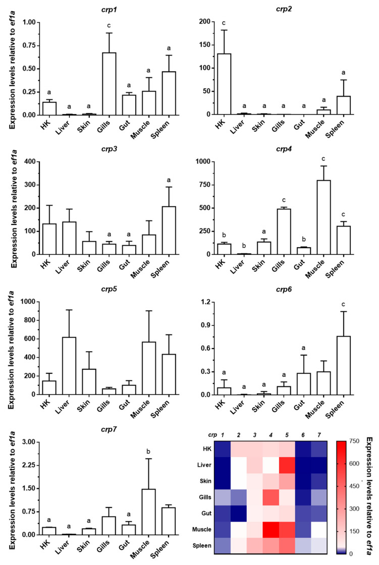
Figure 1 Gene expression analysis of crp1–7 in tissues of healthy zebrafish. The expression of crp1–7 was determined by RT-qPCR by using specific primers for each isoform (Table S1). ef1a mRNA was used as the endogenous control to normalize data, which are represented as the mean relative expression level × 103 ± SD of four different individuals. Statistical differences (p < 0.05, one-way ANOVA) between tissues are represented by: a (different from up to 1–2 tissues), b (different from up to 3–4 tissues), and c (different from up to 5–6 tissues). Data in bar graphs are summarized in a final double gradient colormap (descending blue gradient for values from 0 to 1 and ascending red gradient from values from 1 to ≥750).