
|
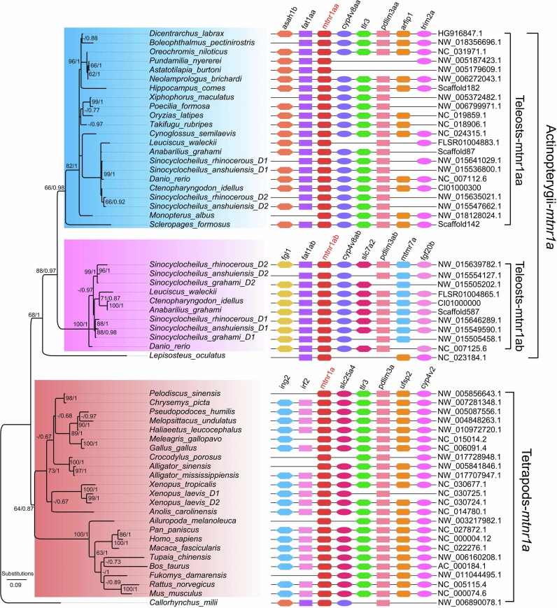
Fig. 2A Fig. 2. Melatonin phylogenetic trees and genome synteny of melatonin receptors in vertebrates. (Fig. 2A) The phylogenetic tree was constructed on the basis of 57 MTNR1A protein sequences (the left part), and the synteny data of mtnr1a (the right part) were presented for validation. (Fig. 2B) The phylogenetic tree was constructed on the basis of 59 MTNR1B protein sequences (the left part), and the synteny data of mtnr1b (the right part) were presented for confirmation. (Fig. 2C) The phylogenetic tree was constructed on the basis of 48 MTNR1C protein sequences (the left part), with the synteny data of mtnr1c (the right part) for validation. Numbers on the branches from left to right are bootstrap values generated in the PhyML reconstruction and the Bayesian posterior probabilities obtained by the Bayesian inference, respectively. Those bootstrap values under 60% and posterior probabilities less than 0.60 are not shown.
Reprinted from Gene, 769, Li, Y., Lv, Y., Bian, C., You, X., Shi, Q., Molecular evolution of melatonin receptor genes (mtnr) in vertebrates and its shedding light on mtnr1c, 145256, Copyright (2020) with permission from Elsevier. Full text @ Gene