IMAGE

Fig 3

ID
ZDB-IMAGE-210224-26
Source
Figures for Bohns et al., 2020
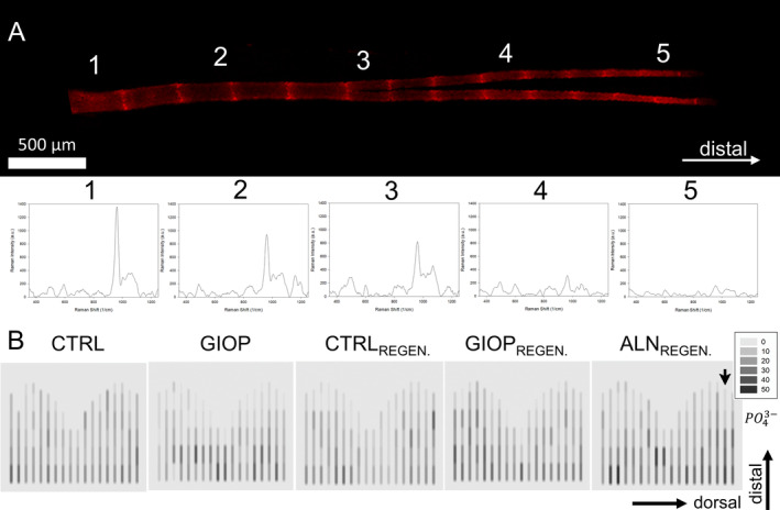
Image
Figure Caption

Fig 3

Montage containing a representative zebrafish caudal fin bony ray and the Raman spectra obtained. (A) Representation of the 17th lepidotrichia (dorsal region of the fin) of the ALNREGEN group isolated from the fluorescence images obtained in confocal microscope; five different spots of interest along the growth direction were measured (A1–5) and the respective Raman peaks were obtained for each region. (B) Heat color plots representing the total area below the curve for the phosphate peak of ~962 cm−1; the black arrow indicates the bony ray represented in the Raman spectra shifts above. The legend box shows the total values of the areas divided by 103. The darker the spots, the higher the mineral intensity of the signal.

Acknowledgments
This image is the copyrighted work of the attributed author or publisher, and ZFIN has permission only to display this image to its users. Additional permissions should be obtained from the applicable author or publisher of the image. Full text @ JBMR Plus