
Figure 6—figure supplement 1.
- ID
- ZDB-IMAGE-210220-36
- Publication
- Fontenas et al., 2021 - Spinal cord precursors utilize neural crest cell mechanisms to generate hybrid peripheral myelinating glia
- All Figures
- Figures for Fontenas et al., 2021
|
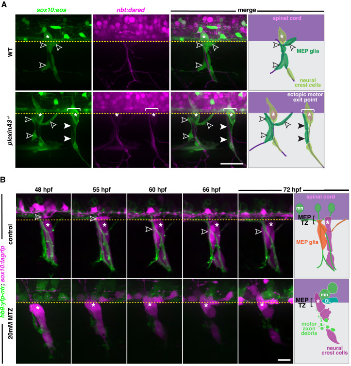
Figure 6—figure supplement 1. (A) Lateral view of sox10:eos;nbt:dsred WT control and plexinA3-/- siblings at 3 dpf showing MEP glia along both motor nerve axons (outlined arrowheads) and ectopic motor nerve axons (arrowheads). Brackets denote ectopic motor exit points. (B) Lateral views of sox10:tagrfp;hb9:yfp-ntr larvae from 48 to 72 hpf, treated with either 2% DMSO or 20 mM metronidazole(MTZ)/2% DMSO from 9 to 72 hpf, showing sox10+ MEP glia (white outlined arrowheads) exit the spinal cord and migrate onto healthy motor axons in the control embryo. In the MTZ-treated larvae, MEP glia did not exit the spinal cord. Asterisks denote the DRG and yellow dashed lines denote the edge of the spinal cord. Scale bar, (A) 25 µm, (B) 20 µm.