
Figure 2—figure supplement 1.
- ID
- ZDB-IMAGE-210220-28
- Publication
- Fontenas et al., 2021 - Spinal cord precursors utilize neural crest cell mechanisms to generate hybrid peripheral myelinating glia
- All Figures
- Figures for Fontenas et al., 2021
|
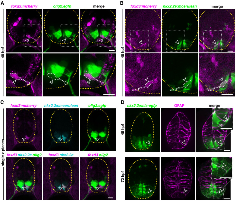
Figure 2—figure supplement 1. (A) Transverse section of a foxd3:mcherry;olig2:egfp embryo at 48 hpf showing a foxd3+/olig2+ MEP glial cell (outlined arrowhead) ventral to the olig2 pMN domain in the spinal cord. (B) Transverse section of a foxd3:mcherry;nkx2.2a:mcerulean embryo at 48 hpf showing a foxd3+/nkx2.2a+ MEP glial cell (outlined arrowhead). (C) Transverse section of a foxd3:mcherry;nkx2.2a:mcerulean;olig2:egfp embryo at 48 hpf showing a foxd3+/nkx2.2a+/olig2+ triple positive MEP glial cell (outlined arrowhead) in the p3 domain of the neural tube, just ventral to the pMN domain. (D) Transverse section of a nkx2.2a:nls-egfp embryo showing nkx2.2a+/GFAP+ radial glia (outlined arrowhead) at 48 hpf (top panel) and 72 hpf (bottom panel). Yellow dashed lines outline the edge of the spinal cord. Top right corner white boxes show higher magnification of bottom white boxes. Scale bar, (A–D) 10 µm.