Image
Figure Caption

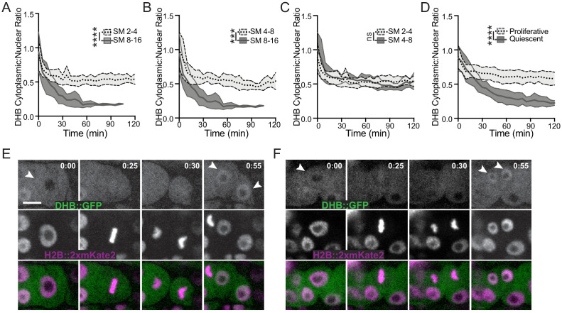
Figure 2—figure supplement 1. CDK sensor can detect differences in proliferation potential of the SM and uterine postembryonic blast lineages.

(A) Cells exit the first and second pre-terminal SM divisions at comparable CDKinc states. (B, C) Cells exit the third and terminal SM division in a CDKlow state that is distinct from that of the earlier divisions. (D) DHB ratios in SS/VU cells, comparing proliferative cells, which exit in a CDKinc state, and quiescent cells, which exit in a CDKlow state (n ≥ 10 each). (E, F) Stills of time-lapse Videos (see Figure 2—video 1) showing CDK sensor localization in cycling SS (E) and quiescent sheath cells (F). Scale bar = 5 μm.

Acknowledgments
This image is the copyrighted work of the attributed author or publisher, and ZFIN has permission only to display this image to its users. Additional permissions should be obtained from the applicable author or publisher of the image. Full text @ Elife