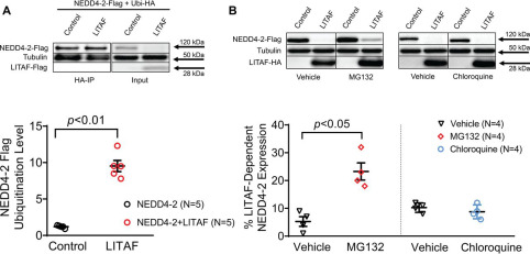
Fig. 6 Figure 6. LITAF causes ubiquitination and subsequent proteasomal degradation of NEDD4-2 in HEK cells. A, IP of lysates from HEK cells transfected with plasmids for FLAG-tagged NEDD4-2, HA-tagged ubiquitin, FLAG-tagged LITAF, or control plasmid for 48 h was performed with anti-HA antiserum. A representative immunoblot shows levels of ubiquitinated NEDD4-2–FLAG and tubulin and input levels of NEDD4-2–FLAG, LITAF-FLAG, or tubulin. Relative NEDD4-2–FLAG ubiquitination levels as calculated by the ratio of ubiquitinated NEDD4-2 normalized to ubiquitinated tubulin and total NEDD4-2 normalized to total ubiquitin levels (n = 5; means ± S.E.). In the scatter plot, averaged values for the five independent experiments together with the means and S.E. values are shown. Bottom panel, Student's t test, p < 0.01. B, LITAF-mediated degradation of NEDD4-2 through proteasomes. HEK cells were transfected with plasmids for NEDD4-2–FLAG, LITAF-HA or control plasmid for 24 h and then treated with vehicle (▿), 5 μm MG132, or 10 μm chloroquine for 24 h. Top panel, representative Western blots show total expression of NEDD4-2–FLAG, LITAF-HA, and tubulin of treated cells. Bottom panel, respective relative expression levels (means ± S.E.) of total NEDD4-2 normalized to tubulin levels (n = 4, n = 2, p < 0.05).
Image
Figure Caption
Acknowledgments
This image is the copyrighted work of the attributed author or publisher, and
ZFIN has permission only to display this image to its users.
Additional permissions should be obtained from the applicable author or publisher of the image.
Full text @ J. Biol. Chem.

