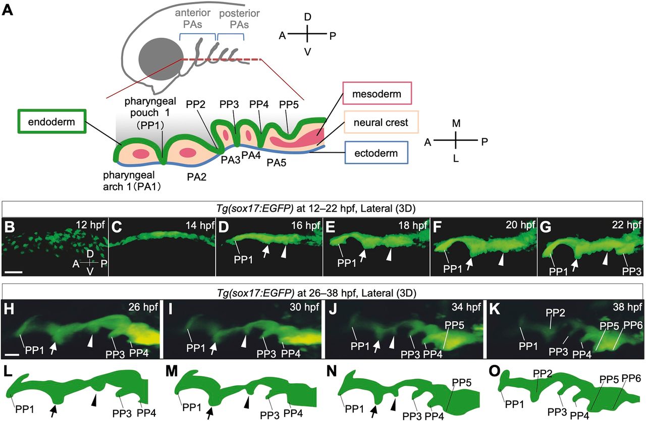
Fig. 1 Time-lapse observations of pharyngeal endoderm during PP segmentation in Tg(sox17:EGFP) zebrafish embryos. (A) Schematic of the bilateral arrangement of PAs in the ventral region of the head. (B-K) Time-lapse analysis of the pharyngeal endoderm of Tg(sox17:EGFP) zebrafish from 12 hpf to 22 hpf (B-G, Movie 1) and from 26 hpf to 38 hpf (H-K, Movie 2). Rostral (arrows) and caudal (arrowheads) bulges appeared posterior to PP1 and gradually fused to form PP2. (L-O) Schematic illustrations of the shape of the lateral pharyngeal endoderm in H-K, respectively. A, anterior; D, dorsal; L, lateral; M, medial; P, posterior; PA1-5, the first to fifth pharyngeal arch; PP1-6, the first to sixth pharyngeal pouches; V, ventral. Scale bars: 50 μm (B) and 20 μm (H).
Image
Figure Caption
Figure Data
Acknowledgments
This image is the copyrighted work of the attributed author or publisher, and
ZFIN has permission only to display this image to its users.
Additional permissions should be obtained from the applicable author or publisher of the image.
Full text @ Development

