
Fig. 4
- ID
- ZDB-IMAGE-210216-72
- Publication
- Rieckhoff et al., 2020 - Spindle Scaling Is Governed by Cell Boundary Regulation of Microtubule Nucleation
- All Figures
- Figures for Rieckhoff et al., 2020
|
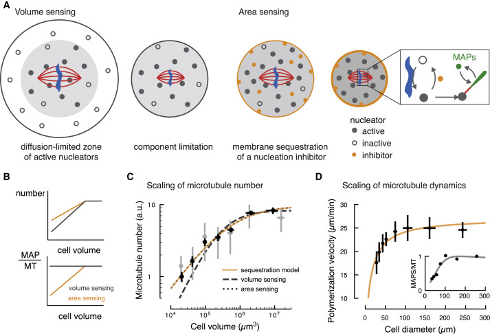
Fig. 4 Figure 4. A Theoretical Model Based on Membrane Sequestration of a Microtubule Nucleation Inhibitor Accounts for Spindle Scaling (A) Left: schematic representation of the limiting component model (“volume sensing”). In large cells, the activation of nucleators (shown as filled circles) near chromosomes is limited by diffusion. As cell size decreases below the diffusion-limited volume, the number of nucleators scale with cell volume. Right: schematic representation of the membrane sequestration model (“area sensing”). An inhibitor of microtubule nucleation (orange circle) is adsorbed at the cell boundary. Inset: activation of nucleators occurs around DNA (in blue), while an inhibitor of microtubule nucleation (orange) triggers its inactivation. Active nucleators can generate new microtubules (red line), to which microtubule-associated proteins (MAPs, in green) can bind. (B) Prediction of how volume sensing and area sensing affect microtubule number and dynamics. If both microtubule number and factors promoting microtubule growth (MAPs) scale equally with cell volume, their ratio remains constant. Only if they scale differently with cell size, their ratio changes and microtubule dynamics may change. (C) Microtubule number is sensitive to the surface area of the cell and it is accounted by the membrane sequestration model (see Figure 3A and STAR Methods, orange line). Data taken from Figure 3A. (D) The membrane sequestration model (orange line) captures the change in polymerization velocity with cell size (see Figure 2A and STAR Methods, mean ± SD). Inset: the number of MAPs per microtubule decreases with decreasing cell diameter. MT, microtubule.