IMAGE

Fig 7

ID
ZDB-IMAGE-210128-52
Source
Figures for Lin et al., 2020
Image
Figure Caption

Fig 7 Functional validation of four asymmetrically localized transcripts encoding transcription factors.

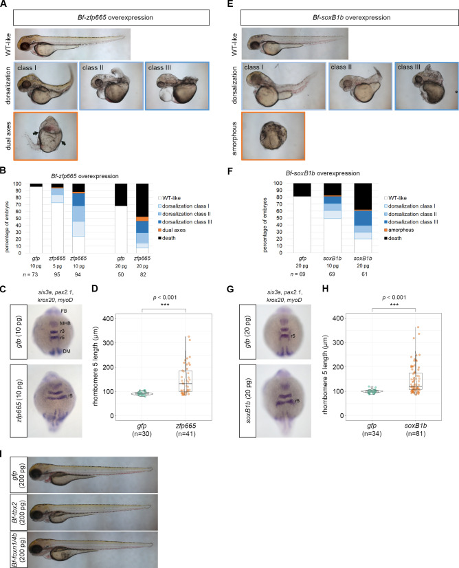
(A) Phenotypes of the 48 hpf zebrafish embryos injected with amphioxus zfp665 mRNA. (B) Percentages of 48 hpf zebrafish embryos with the indicated phenotypes following injection of gfp (control) or amphioxus zfp665 mRNA. (C) WMISH of pax2.1, krox20, myoD and six3a in gfp (control) or amphioxus zfp665 mRNA-injected zebrafish embryos at the 10 somite stage. (D) Statistical analysis of the lengths of rhombomeres 5 in gfp and zfp665 mRNA injected zebrafish embryos at the 10 somite stage. (E) Phenotype of the 48 hpf zebrafish embryos injected with amphioxus soxB1b mRNA. (F) Percentages of 48 hpf zebrafish embryos with the indicated phenotypes after injection of gfp (control) or amphioxus soxB1b mRNA. (G) WMISH of pax2.1, krox20, myoD and six3a in gfp (control) or amphioxus soxB1b mRNA-injected zebrafish embryos at the 10 somite stage. (H) Statistical analysis of the lengths of rhombomeres 5 in gfp and soxB1b mRNA-injected zebrafish embryos at the 10 somite stage. (I) Embryos injected with amphioxus tbx2 or foxn1/4b mRNA did not show any detectable axial defects compared to control embryos at 72 hpf. DM, dorsal mesoderm; FB, forebrain; MHB, midbrain-hindbrain boundary; r3, rhombomere 3; r5, rhombomere 5. Statistical significance was determined by Student’s t-test. ***p < 0.001. Underlying data are available in S1 Data.

Acknowledgments
This image is the copyrighted work of the attributed author or publisher, and ZFIN has permission only to display this image to its users. Additional permissions should be obtained from the applicable author or publisher of the image. Full text @ PLoS Genet.