
Figure 2
- ID
- ZDB-IMAGE-210128-106
- Publication
- Takaki et al., 2020 - Schistosoma mansoni Eggs Modulate the Timing of Granuloma Formation to Promote Transmission
- All Figures
- Figures for Takaki et al., 2020
|
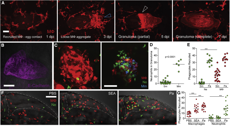
Figure 2
(A) Time-lapse microscopy of egg monitored at 2-day intervals from 1–7 dpi showing in the four panels, respectively, sequential macrophage recruitment, aggregation (blue arrowhead), formation of the partial granuloma (white arrowhead), and its expansion to encase the egg. Scale bar, 25 μm.
(B) Epithelioid granuloma immunostained using E-cadherin antibody. Scale bar, 50 μm.
(C) Confocal images of granulomas in representative transgenic zebrafish larvae with red-fluorescent macrophages (MΦ) and green-fluorescent neutrophils (Ne) at 5 dpi with
(D) Quantification of neutrophils recruited to Sm and Mm granulomas.
(E) Quantification of phagocytes recruited to Sm and
(F) Confocal images of HBV of representative larvae showing phagocyte recruitment at 6 h post-injection with phosphate buffered saline (PBS) (left),
(G) Quantification of phagocytes recruited to Sm and