IMAGE

Figure 3

ID
ZDB-IMAGE-210117-41
Source
Figures for de Latouliere et al., 2021
Image
Figure Caption

Figure 3

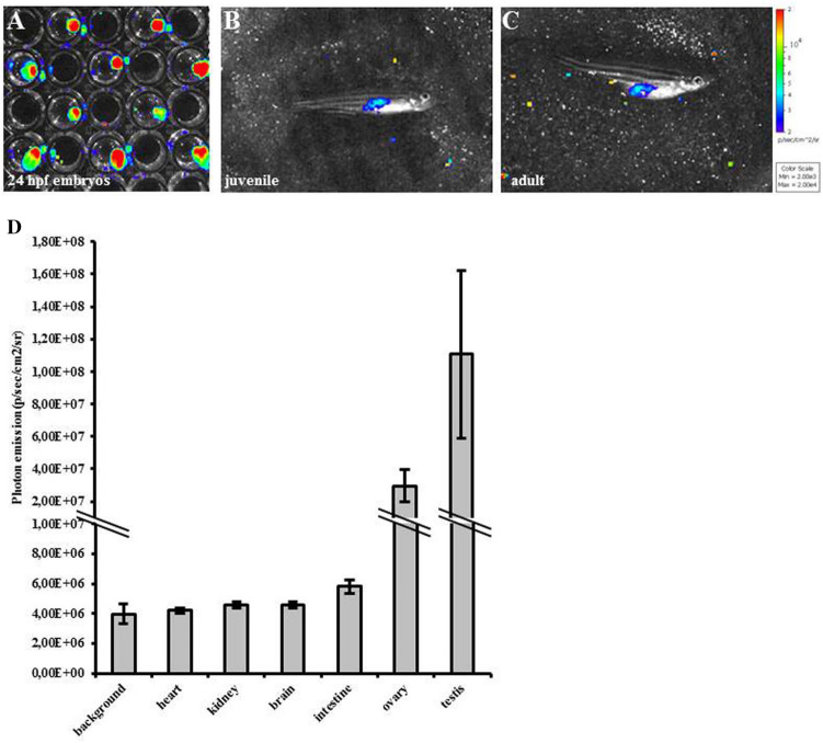
Bioluminescence correlates with proliferation in MITO-Luc/GFP zebrafish line. (A–C) In vivo BLI of representative MITO-Luc/GFP1 zebrafish embryos (1 embryo for each well) (A), juvenile (B) and adult (C) stages. Light emitted from the animal appears in pseudocolor scaling. (D) Photon emission quantification of organs extracted from adult zebrafish. The error bars are mean standard deviations from six samples for each organ.

Acknowledgments
This image is the copyrighted work of the attributed author or publisher, and ZFIN has permission only to display this image to its users. Additional permissions should be obtained from the applicable author or publisher of the image. Full text @ Sci. Rep.