IMAGE

Fig. 7

ID
ZDB-IMAGE-210117-34
Source
Figures for Kuo et al., 2021
Image
Figure Caption

Fig. 7

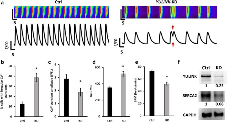
Assessment of arrhythmia and irregular Ca2+ regulation in YULINK KD iPSC-derived human cardiomyocytes. a Representative Ca2+ scans and spontaneous Ca2+ transient in control and YULINK KD iPSC-derived human cardiomyocytes. Red arrows indicate abnormal arrhythmia-like Ca2+ waveforms observed in YULINK KD cardiomyocytes. b Percentages of the irregular Ca2+ transients in control and YULINK KD iPSC-derived human cardiomyocytes (n = 40). c Bar graph comparison of Ca2+ transient amplitude of human iPSC-derived cardiomyocytes for control and YULINK KD confirmed that YULINK KD human cardiomyocytes exhibited a lower Ca2+ transient amplitude (n = 40). d The time constant for Ca2+ decay (Tau) of human iPSC-derived cardiomyocytes for control and YULINK KD showed that the time constant for Ca2+ decay was significantly larger in YULINK KD than in control iPSC-derived human cardiomyocytes (n = 40). e Quantification of spontaneous beating rate of control and YULINK KD human cardiomyocytes (n = 40). (*p < 0.05, Student’s t test). f Human cardiomyocytes were subjected to YULINK KD using YULINK-shRNA and the effects on YULINK and SERCA2 expression were assayed by Western blot

Acknowledgments
This image is the copyrighted work of the attributed author or publisher, and ZFIN has permission only to display this image to its users. Additional permissions should be obtained from the applicable author or publisher of the image. Full text @ J. Biomed. Sci.