
Fig 1
- ID
- ZDB-IMAGE-210113-118
- Publication
- Nunley et al., 2020 - Defect patterns on the curved surface of fish retinae suggest a mechanism of cone mosaic formation
- All Figures
- Figures for Nunley et al., 2020
|
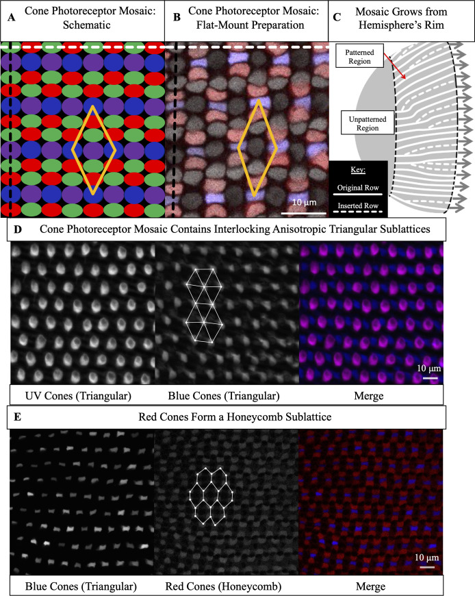
Fig 1
(A) Schematic of cone photoreceptors (colored by subtype) in apical plane of zebrafish retina. The ‘unit cell’ (yellow parallelogram) contains one UV cone, one Blue cone, two Green cones, and two Red cones. White dashed line: ‘row’ axis. Black dashed line: ‘column’ axis. (B) Cone mosaic from flat-mount retinal preparation of an adult, triple transgenic fish, Tg[