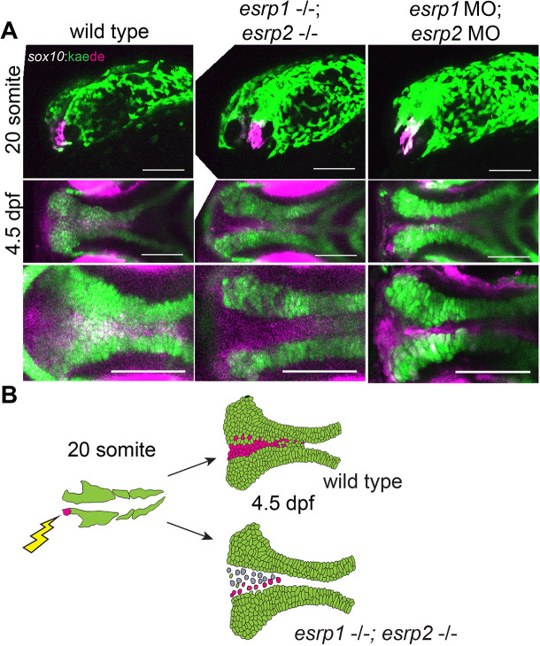
esrp1/2 null cranial neural crest cells(CNCCs)migrate to the ANC but do not differentiate to chondrocytes. (A) Lineage tracing of WT or esrp1/2 morphant zebrafish embryos using the Tg(sox10:kaede) line, native Kaede fluorescence is shown in green, and photo-converted Kaede is shown in magenta. Sagittal and horizontal views of zebrafish embryos at 19 hpf and 4.5 dpf, respectively. The anteriormost neural crest frontonasal prominence (FNP) progenitors were photoconverted at 19 hpf. At 4.5 dpf, the WT signal tracks to the medial portion of the ANC. Both the esrp1/esrp2 double CRISPR mutants and esrp1/2 morphants exhibit a cleft in the ANC with absence of a portion of sox10+ cells in the medial portion of the ANC, but the labeled CNCCs representing FNP progenitors did reach and populate the entire length of the ANC. (B) Illustrative summary of lineage tracing results showing that photo-converted anteriormost CNCCs contributing to FNP do migrate into the ANC in esrp1/2 mutant embryos, but a cleft forms at the juxtaposition of the FNP-derived median element and the maxillary-derived lateral element. Scale bars: 150 µm.
This image is the copyrighted work of the attributed author or publisher, and
ZFIN has permission only to display this image to its users.
Additional permissions should be obtained from the applicable author or publisher of the image.
Full text @ Development
Your Input Welcome
Thank you for submitting comments. Your input has been emailed to ZFIN curators who may contact you if
additional information is required.
Oops. Something went wrong. Please try again later.