
FIGURE 7
- ID
- ZDB-IMAGE-210103-29
- Publication
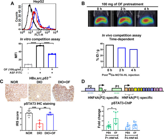
- Wu et al., 2020 - Low molecular weight fucoidan inhibits hepatocarcinogenesis and nonalcoholic fatty liver disease in zebrafish via ASGR/STAT3/HNF4A signaling
- All Figures
- Figures for Wu et al., 2020
|
FIGURE 7
Oligo‐fucoidan binds to ASGR1/2 in hepatoma cells, enhances pSTAT3 in hepatic tissues of [